Journal of caring sciences.15(1) :61-64.
doi: 10.34172/jcs.026.33750
Case Report
Iatrogenic Fracture of a Peripheral Intravenous Cannula: A Case Report
Anandaperumal Pachiyappan Conceptualization, Data curation, Formal analysis, Investigation, Methodology, Project administration, Resources, Project administration, Supervision, Validation, Visualization, Writing – original draft, Writing – review & editing, 1 
Amirthavalli Athimulam Conceptualization, Data curation, Formal analysis, Investigation, Methodology, Project administration, Resources, Project administration, Supervision, Validation, Visualization, Writing – original draft, Writing – review & editing, 1
Kalayarasan Raja Conceptualization, Data curation, Formal analysis, Investigation, Methodology, Project administration, Resources, Project administration, Supervision, Validation, Visualization, Writing – original draft, Writing – review & editing, 2
Lakshmi Ramamoorthy Methodology, Project administration, Resources, Project administration, Supervision, Validation, Visualization, Writing – original draft, Writing – review & editing, 3, *
Author information:
1Liver Transplant ICU, JIPMER,Puducherry
2Department of Surgical Gastroenterology, JIPMER
3College of Nursing, JIPMER, Puducherry-06, India
Abstract
Introduction:
Peripheral intravenous cannulation is a routine invasive procedure widely used in healthcare settings. While it is typically associated with minimal risk, uncommon complications such as cannula fracture and intravascular migration may occur. They can result in significant morbidity if diagnosis and intervention are delayed.
Case report:
We describe an adult patient who developed an iatrogenic fracture of a peripheral intravenous cannula, with proximal displacement of the fractured segment into the brachial vein. Ultrasonography was employed to identify and localize the intravascular foreign body. A catheter fragment measuring approximately 2.5 cm was surgically removed via venotomy performed about 5 cm proximal to the initial cannulation site. The patient recovered uneventfully, with no postoperative complications.
Conclusion:
Although rare, fracture and migration of peripheral intravenous cannulas constitute a serious iatrogenic event. Prompt imaging, particularly with ultrasound, is essential for accurate detection and localization. This case emphasizes the need for meticulous cannula insertion and removal practices, along with heightened clinical vigilance, to improve patient safety and prevent avoidable complications.
Keywords: Peripheral intravenous catheter, Iatrogenic injury, Foreign body, Catheter fracture
Copyright and License Information
© 2025 The Author(s).
This work is published by Journal of Caring Sciences as an open access article distributed under the terms of the Creative Commons Attribution Non-Commercial License (
http://creativecommons.org/licenses/by-nc/4.0/). Non-commercial uses of the work are permitted, provided the original work is properly cited.
Funding Statement
None.
Introduction
Peripheral intravenous catheters (PIVCs) are the most commonly used invasive devices in hospitals worldwide, with billions inserted annually for the administration of fluids, medications, blood products, and parenteral nutrition.1,2 Although considered routine, PIVCs are associated with high rates of non-infectious complications and failure. Extensive systematic reviews estimate all-cause PIVC failure at around 36–40%, mainly due to phlebitis, infiltration/extravasation, occlusion, leakage, and dislodgement.3,4 Individual cohort studies frequently report phlebitis rates of 9–44% and infiltration rates of 10–16%. These events interrupt therapy, increase the number of procedures, prolong hospital stays, and increase costs.
Historically, guidelines from the CDC and the Infusion Nurses Society recommended routine replacement every 72–96 hours in adults to reduce the risk of phlebitis and infection.5 However, several randomised trials and meta-analyses now show that clinically indicated replacement (removal for signs of phlebitis, infiltration, occlusion, infection, or end of therapy) is not inferior to routine 72–96-hour resite for phlebitis or bloodstream infection, while reducing the number of cannulas and costs.6,7 A large equivalence trial in Australia (3,283 patients) found identical phlebitis incidence (7%) between clinically indicated and third-daily replacement.5 Similar results have been reported in Brazil and China, with no increase in phlebitis, infection, or mortality, although some studies observed slightly higher rates of clinically indicated replacement.8,9 Contemporary evidence-based recommendations therefore favour replacement based on clinical indication rather than fixed time intervals in adult patients, provided meticulous insertion technique and ongoing site surveillance are in place.8
Despite adherence to such guidance, mechanical complications remain a significant problem. Large datasets indicate that overall PIVC failure occurs in approximately one-third of devices, with female sex, older age, high-risk infusates (e.g., IV antibiotics, vasoactive or irritant drugs), small-gauge catheters, joint sites (hand, wrist, antecubital fossa), and longer dwell times as significant risk factors. Fracture of the peripheral cannula, with intravascular migration of a plastic fragment, is exceptionally rare and not captured in large epidemiological series, being described almost exclusively in case reports. Reinsertions of the metal stylet into a partially or fully advanced plastic catheter, forceful manipulation, repeated attempts with the same device, and prolonged dwell time at areas of flexion are repeatedly implicated as preventable technical causes. 10,11
Although uncommon, embolised catheter fragments may lead to severe outcomes, including sepsis, endocarditis, vascular or cardiac perforation, thrombosis, arrhythmia, pulmonary embolism, myocardial infarction, and even death.10 Early recognition, prompt imaging to localise the fragment, and timely surgical or endovascular retrieval are therefore crucial.12,13. This case report describes a rare complication of peripheral intravenous cannulation and highlights preventive strategies to enhance patient safety.
Research Highlights
What is the current knowledge?
-
Peripheral intravenous cannula fracture is a rare iatrogenic complication, most often occurring during insertion or manipulation.
-
Migration of the fractured segment into the central circulation may result in serious, life-threatening complications.
-
Endovascular retrieval is considered the gold-standard treatment for centrally embolized fragments.
What is new here?
-
Cannula fracture occurred during a routine discharge procedure, highlighting that complications may arise at any stage of care.
-
Immediate application of a proximal tourniquet prevented embolization of the fragment.
-
The fragment lodged in a peripheral vein, making surgical exploration under local anesthesia a safe and effective primary approach.
Presentation of the Case
A 23-year-old male presented with abdominal pain and was diagnosed with pancreatitis. He subsequently underwent robot-assisted bilateral thoracic splanchnic neurectomy involving the bile duct and duodenum, with preservation of the pancreatic head. During his postoperative hospital stay, a 20 Gauge peripheral intravenous (IV) cannula was in situ at the cephalic vein of the forearm for routine medication administration. The postoperative course was uneventful, and discharge was planned for 1 week postoperatively.
At the time of discharge, during routine removal of the IV cannula, it was observed that a portion of the plastic catheter had fractured and was retained at the cannulation site. The incident was immediately reported to the nurse in charge and the duty medical officer. A tourniquet was promptly applied proximal to the insertion site to prevent proximal migration of the fragment.
Cardiothoracic and vascular surgery consultations were obtained without delay, and a bedside ultrasonography (USG) examination was performed, which suggested the presence of a retained intravascular foreign body. The location of the fractured cannula fragment was subsequently confirmed by imaging. Based on these findings, a general surgery consultation was sought, and surgical exploration under local anesthesia was advised for foreign body removal.
After detailed preoperative counseling regarding the potential risks and benefits, informed consent was obtained from the patient. Under sterile conditions, local anesthesia was achieved by infiltrating 2% lignocaine over the marked site and surrounding area. A 2 cm longitudinal incision was made over the course of the cephalic vein using a size 15 blade. The wound was explored, and the cephalic vein was identified. As the cannula fragment was not immediately visualized or palpated, the incision was extended proximally by an additional 2 cm along the vein’s course. The retained cannula fragment was then palpated, and a partial venotomy was performed, allowing successful extraction of the fractured component from the vein (Figure 1).
Discussion
Fracture of a peripheral intravenous (IV) cannula is an infrequently reported complication, particularly when compared with fractures involving central venous access devices.⁵ Published literature indicates that the most likely contributing factor is reinsertion of the stylet into a plastic catheter that has already been advanced into the vein. When the catheter follows a curved venous path or is inserted at an oblique angle, reinserting the stylet can compromise the catheter wall, resulting in partial or complete structural failure.14,15
In the present case, it is postulated that partial damage to the catheter occurred during cannulation. At the time of catheter removal, the weakened segment may have become embedded in the vessel intima, and continued traction likely led to complete separation of the distal fragment, leaving it intravascularly retained. This sequence of events highlights the importance of nursing technique and vigilance, as nurses are primarily responsible for peripheral IV insertion, maintenance, and removal in routine clinical care.
Preventive strategies are therefore central to nursing practice. Existing standards and best-practice recommendations advise against reinserting the stylet once the plastic catheter has entered the vein, discourage multiple attempts with the same cannula, and emphasize appropriate vein selection and secure catheter stabilization to limit mechanical stress.8,16 Regular assessment of catheter necessity and timely removal when IV access is no longer required are also recommended.17,18 Any recommendations regarding limiting cannulation attempts should be applied judiciously and in accordance with institutional policies and clinical judgment.5,9,19
Early identification of complications is equally crucial. Nurses should carefully inspect the catheter upon removal to ensure that it is intact. Failure to detect a fractured cannula promptly may result in delayed diagnosis and increase the risk of complications such as proximal migration, infection, vascular injury, and extended hospitalization.20,21 These complications may also increase healthcare costs due to the need for imaging studies and invasive retrieval procedures.10,22
Although several endovascular and surgical techniques—such as snare devices, balloon catheters, and retrieval forceps—have been described for removing intravascular foreign bodies,prevention remains the most effective approach.23,24 Ongoing education, competency-based training, and strict adherence to established IV therapy guidelines are essential nursing-led measures to enhance patient safety. Additionally, clear communication with patients regarding IV therapy and prompt management of adverse events may help reduce dissatisfaction and medicolegal concerns.
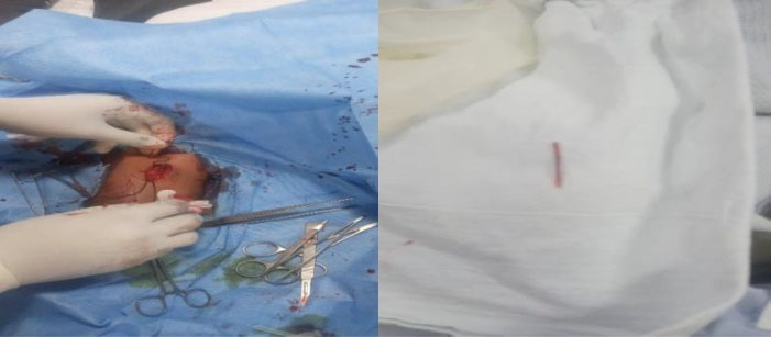
Figure 1.
Surgical exploration of a fragmented peripherally inserted intravenous cannula
.
Surgical exploration of a fragmented peripherally inserted intravenous cannula
Conclusion
While a peripheral IV cannula fracture is rare, it is largely avoidable. Emphasizing safe cannulation practices, avoiding stylet reinsertion, ensuring careful monitoring, and verifying catheter integrity at removal—consistent with established guidelines—can substantially reduce the risk of this preventable complication. A careful history and physical examination should be performed to diagnose a broken cannula in a peripheral vein. Before retrieval, imaging should be performed to localize the broken fragment, and tourniquets should be used to prevent distal migration. Given the potential for fatal outcomes from intravascular migration, early exploration should be the priority.
Competing Interests
The Authors declare no conflict of interest.
Data Availability Statement
Data are available upon reasonable request to the corresponding author.
Ethical Approval
The patient provided written informed consent. Written informed consent was obtained from the individual for the publication of any potentially identifiable images or data included in this article.
References
- Shinzato Y, Yasuda H, Moriya T, Taira H, Kishihara Y, Kashiura M. Risk factors of phlebitis in patients admitted to the intensive care unit vary according to the duration of catheter dwelling: A post-hoc analysis of the AMOR-VENUS study. PLoS One 2025; 20(4):e0320583. doi: 10.1371/journal.pone.0320583 [Crossref] [ Google Scholar]
- Marsh N, Larsen EN, Ullman AJ, Mihala G, Cooke M, Chopra V. Peripheral intravenous catheter infection and failure: A systematic review and meta-analysis. Int J Nurs Stud 2024; 151:104673. doi: 10.1016/j.ijnurstu.2023.104673 [Crossref] [ Google Scholar]
- Blanco-Mavillard I, de Pedro-Gómez JE, Rodríguez-Calero MÁ, Bennasar-Veny M, Parra-García G, Fernández-Fernández I. Multimodal intervention for preventing peripheral intravenous catheter failure in adults (PREBACP): a multicentre, cluster-randomised, controlled trial. Lancet Haematol 2021; 8(9):e637-47. doi: 10.1016/S2352-3026(21)00173-7 [Crossref] [ Google Scholar]
- Indarwati F, Mathew S, Munday J, Keogh S. Incidence of peripheral intravenous catheter failure and complications in paediatric patients: Systematic review and meta analysis. Int J Nurs Stud 2020; 102:103488. doi: 10.1016/j.ijnurstu.2019.103488 [Crossref] [ Google Scholar]
- Rickard CM, Webster J, Wallis MC, Marsh N, McGrail MR, French V. Routine versus clinically indicated replacement of peripheral intravenous catheters: a randomised controlled equivalence trial. Lancet 2012; 380(9847):1066-74. doi: 10.1016/s0140-6736(12)61082-4 [Crossref] [ Google Scholar]
- Rickard CM, McCann D, Munnings J, McGrail MR. Routine resite of peripheral intravenous devices every 3 days did not reduce complications compared with clinically indicated resite: a randomised controlled trial. BMC Med 2010; 8:53. doi: 10.1186/1741-7015-8-53 [Crossref] [ Google Scholar]
- Li J, Ding Y, Lu Q, Jin S, Zhang P, Jiang Z. Routine replacement versus replacement as clinical indicated of peripheral intravenous catheters: A multisite randomised controlled trial. J Clin Nurs 2022; 31(19-20):2959-70. doi: 10.1111/jocn.16129 [Crossref] [ Google Scholar]
- Zanella MC, Catho G, Jackson H, Lotfinejad N, Sauvan V, Chraiti MN. Dwell Time and Risk of Bloodstream Infection With Peripheral Intravenous Catheters. JAMA Netw Open 2025; 8(4):e257202. doi: 10.1001/jamanetworkopen.2025.7202 [Crossref] [ Google Scholar]
- Drugeon B, Rickard CM, Peters N, Chopra V, Mimoz O. Procedural and Therapy Peripheral Intravenous Catheters: A Framework for Safety, Quality and Patient-Centred Care. J Adv Nurs 2025. 10.1111/jan.70347
- Simin D, Milutinović D, Turkulov V, Brkić S. Incidence, severity and risk factors of peripheral intravenous cannula-induced complications: An observational prospective study. J Clin Nurs 2019; 28(9-10):1585-99. doi: 10.1111/jocn.14760 [Crossref] [ Google Scholar]
- Yasuda H, Rickard CM, Marsh N, Yamamoto R, Kotani Y, Kishihara Y. Risk factors for peripheral intravascular catheter-related phlebitis in critically ill patients: analysis of 3429 catheters from 23 Japanese intensive care units. Ann Intensive Care 2022; 12(1):33. doi: 10.1186/s13613-022-01009-5 [Crossref] [ Google Scholar]
- Alexandrou E, Ray-Barruel G, Carr PJ, Frost SA, Inwood S, Higgins N. Use of Short Peripheral Intravenous Catheters: Characteristics, Management, and Outcomes Worldwide. J Hosp Med 2018; 13(5):E1-E7. doi: 10.12788/jhm.3039 [Crossref] [ Google Scholar]
- Blanco-Mavillard I, de Pedro-Gómez JE, Rodríguez-Calero M, Bennasar-Veny M, Parra-García G, Fernández-Fernández I. Multimodal intervention for preventing peripheral intravenous catheter failure in adults (PREBACP): a multicentre, cluster-randomised, controlled trial. Lancet Haematol 2021; 8(9):e637-e47. doi: 10.1016/s2352-3026(21)00206-4 [Crossref] [ Google Scholar]
- Blanco-Mavillard I, de Pedro-Gómez JE, Rodríguez-Calero MÁ, Bennasar-Veny M, Parra-García G, Fernández-Fernández I. Multimodal intervention for preventing peripheral intravenous catheter failure in adults (PREBACP): a multicentre, cluster-randomised, controlled trial. The Lancet Haematology 2021; 8(9):e637-e47. [ Google Scholar]
- World Health Organization. Guidelines on the prevention of bloodstream infections and other infections associated with intravascular catheters: Part I – Peripheral catheters. World Health Organization; 2024. Accessed February 13, 2025. Available from:https://www.who.int/publications/i/item/9789240093829.
- Mimoz O, Debonne A, Glanard A, Keita Perse O, Lucet JC. Best practice in the use of peripheral venous catheters: A consensus from French experts. Infect Dis Now 2024; 54(5):104923. doi: 10.1016/j.idnow.2024.104923 [Crossref] [ Google Scholar]
- Amble K, Skjelbreid IB, Eide GE, Muri S, Høvik LH, Reime MH. Implementation of an Infection Prevention Care Bundle for Peripheral Intravenous Catheters (PIVCs): A Quality Improvement Study to Enhance PIVC Quality and Reduce Complications. Nurs Rep 2025; 15(11):379. doi: 10.3390/nursrep15110379 [Crossref] [ Google Scholar]
- Dessalegn A, Ali MS, Yohannes S, Tamir Y, Mulatu S, Zewdie A. Knowledge, practice and associated factors towards intravenous cannula-related infection prevention among nurses working at Northwest Amhara Regional State Comprehensive Specialized Hospitals, Ethiopia. BMC Nurs 2024; 23(1):168. doi: 10.1186/s12912-024-01737-y [Crossref] [ Google Scholar]
- Helm RE, Klausner JD, Klemperer JD, Flint LM, Huang E. Accepted but unacceptable: peripheral IV catheter failure. J Infus Nurs 2015; 38(3):189-203. doi: 10.1097/nan.0000000000000100 [Crossref] [ Google Scholar]
- Ray-Barruel G, Xu H, Marsh N, Cooke M, Rickard CM. Effectiveness of insertion and maintenance bundles in preventing peripheral intravenous catheter-related complications and bloodstream infection in hospital patients: A systematic review. Infect Dis Health 2019; 24(3):152-68. doi: 10.1016/j.idh.2019.03.001 [Crossref] [ Google Scholar]
- Chopra V, Flanders SA, Saint S, Woller SC, O’Grady NP, Safdar N. The Michigan Appropriateness Guide for Intravenous Catheters (MAGIC): Results From a Multispecialty Panel Using the RAND/UCLA Appropriateness Method. Ann Intern Med 2015; 163(6 Suppl):S1-40. doi: 10.7326/m15-0744 [Crossref] [ Google Scholar]
- Liu C, Chen L, Kong D, Lyu F, Luan L, Yang L. Incidence, risk factors and medical cost of peripheral intravenous catheter-related complications in hospitalised adult patients. J Vasc Access 2022; 23(1):57-66. doi: 10.1177/1129729820978124 [Crossref] [ Google Scholar]
- Intagliata E, Basile F, Vecchio R. Totally implantable catheter migration and its percutaneous retrieval: case report and review of the literature. G Chir 2017; 37(5):211-5. doi: 10.11138/gchir/2016.37.5.211 [Crossref] [ Google Scholar]
- Adeosun PO, Abdulazeez AT, Okeke UI, Ehinmosan OE, Eze EG. CT-guided retrieval of a fractured intravenous cannula in a toddler: A case report. Afr J Emerg Med 2020; 10(4):277-80. doi: 10.1016/j.afjem.2020.06.010 [Crossref] [ Google Scholar]